 English
 Deutsch

Phagograms
Bacteriophages are characterized by a high host specificity towards bacteria. The phagogram, similar to the antibiogram, is used to test the sensitivity of clinical bacterial isolates to the lytic activity of bacteriophages. Phagograms are prepared according to the standardized procedure established at the Eliava Institute, Tbilisi, Georgia.
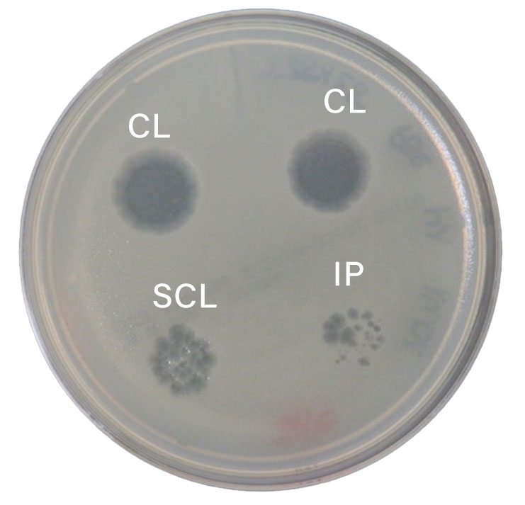
Phagogram on a TS agar plate using the top agar method on P. aeruginosa
The phagogram illustrated shows the in vitro efficacy of different bacteriophage suspensions. The different morphology of the lysis zones can be clearly seen.
CL = “confluent lysis”, clear lysis, one clear lysis zone throughout, positive
SCL = “semi-confluent lysis”, semi-confluent lysis, one clear lysis zone throughout with a faint hazy background, weakly positive
IP = “individual plaques”, individual plaques, weakly positive
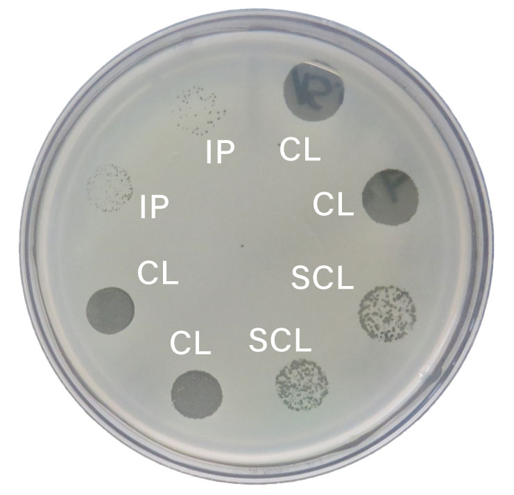
Phagogram on a TS agar plate using the top agar method on S. pseudintermedius
The phagogram illustrated shows the in vitro efficacy of different bacteriophage suspensions. The different morphology of the lysis zones can be clearly seen.
CL = “confluent lysis”, clear lysis, one clear lysis zone throughout, positive
SCL = “semi-confluent lysis”, semi-confluent lysis, one clear lysis zone throughout with a faint hazy background, weakly positive
IP = “individual plaques”, individual plaques, weakly positive
Phagograms
Bacteriophages are characterized by a high host specificity towards bacteria. The phagogram, similar to the antibiogram, is used to test the sensitivity of clinical bacterial isolates to the lytic activity of bacteriophages. Phagograms are prepared according to the standardized procedure established at the Eliava Institute, Tbilisi, Georgia.
Literaturnachweis
-
Chanishvili, N., Myelnikov, D., and Blauvelt, T. K. (2022). Professor Giorgi Eliava and the Eliava Institute of Bacteriophage. Phage (New Rochelle) 3, 71–80. doi: 10.1089/phage.2022.0016
-
E. H. Hankin (1895). Observations on Cholera in India. Ind Med Gaz.
-
Eaton, M. D., and Bayne-Jones, S. (1934). Bacteriophage Therapy. JAMA 103, 1769. doi: 10.1001/jama.1934.72750490003007
-
Felix d’Herelle (1917). On an invisible microbe antagonistic to dysentery bacilli.
-
Florian Georg Leupold (2018). Die Geschichte des VEB Serum-Werk Bernburg von 1954 bis 1990 unter besonderer Berücksichtgung biogener Arzneistoffe.
-
Frederick W. Twort (1915). An investigation on the nature of ultra-microscopic viruses.
-
Hankin, E. H. (1905). On the Epidemiology of Plague. J. Hyg. 5, 48–83. doi: 10.1017/s0022172400002357
-
Krueger, A. P., and Scribner E. Jane (1941). The Bacteriophage. JAMA 116, 2269. doi: 10.1001/jama.1941.62820200013011
-
Ruska, H. (1942). “Morphologische Befunde bei der bakteriophagen Lyse,” in Morphologische Befunde bei der bakteriophagen Lyse (Springer, Berlin, Heidelberg), 345–387.